
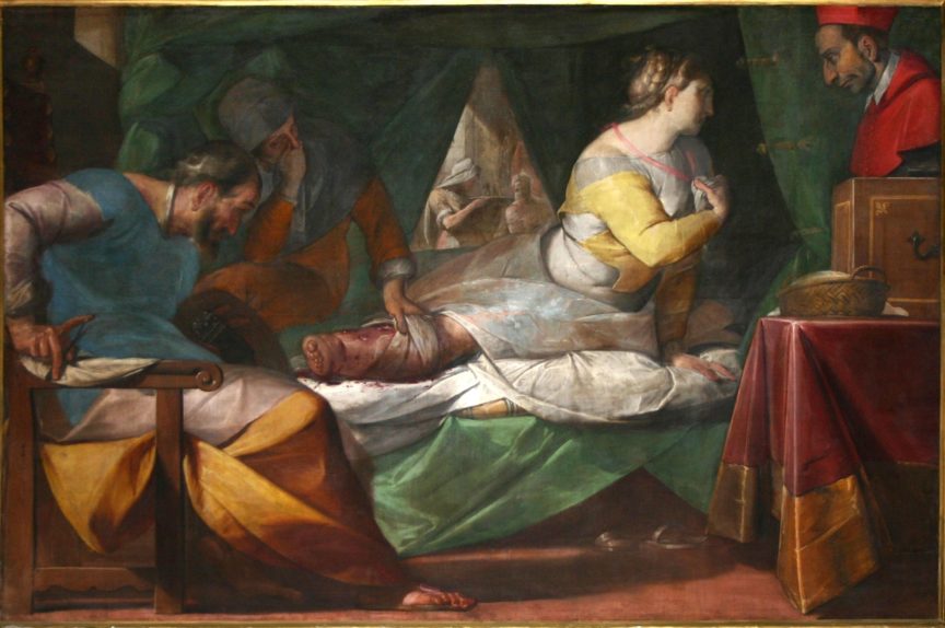

Miracle of Aurelia degli Angeli by Crespi (Click for full size image)
This week’s Spot the Diagnosis! looks at Miracle of Aurelia degli Angeli. Painted on canvas by Giovanni Battista Crespi in 1600, this piece pays homage to the life and miracles of Saint Aurelia degli Angeli. This woman looks to the Saint for aid, while doctors examine her red leg.
While you’re giving it some thought, please consider taking 5 minutes to give us feedback on the “Spot the Diagnosis!” series. From newcomers to loyal fans, we would love to hear about how you use the series and what we can do to improve!
Click here to provide feedback on the “Spot the Diagnosis!” Series!
[bg_faq_start]What is causing the red leg?
The differential diagnosis for this woman includes: chronic stasis, lymphoedema, acute or chronic osteomyelitis, or, most likely, a Strep. pyogenes infection. (Strep causes about 2/3’s of soft tissue infections, and Staph 1/3.)1 Strep causes a variety of infections depending on the layer of tissue to which it invades. Invasion of the keratin layer of the skin results in “crusty” impetigo, a superficial dermis infection is called erysipelas, cellulitis is when it invades the subcutaneous layers, necrotizing fasciitis is when it invades the fascia and a muscle infection for Strep is myonecrosis.2

Infections of the various layers of skin tissue2
| Infection | Layer | Feature |
| Impetigo | Keratin | Crusty lesions |
| Erysipelas | Superficial epidermis | Well-defined borders Bright red colour lesions are raised above the normal skin |
| Cellulitis | Subcutaneous | Pink hue, ill-defined border |
| Necrotizing fasciitis | Fascia + destruction of tissue | Pain out of proportion to how it looks |
| Myonecrosis | Muscle | Purple violaceous bullae Hemorrhage of skin Skin destruction |
The stark swelling, bullae and colour on this woman’s leg point towards erysipelas as the infection ailing her.
What are the clinical manifestations?
Erysipelas is a skin and soft tissue infection which occurs from bacterial entry past the skin barrier into the underlying tissue. The mechanism of bacterial inoculation can vary, with Athlete’s foot being one of the most common causes. Typically found in children and older adults, erysipelas usually presents with symptoms similar to cellulitis – skin erythema, edema, warmth, and fatigue. Most cases are unilateral with a preference for the lower limbs (as illustrated by Crespi).
Although cellulitis and erysipelas can both present similarly, there are certain differences to be aware of. Erysipelas is associated with systemic symptoms prior to onset of erythema such as fever and hypotension, and it is rapidly progressive. Moreover, erysipelas is classically defined by three characteristics: bright red colour; well-defined raised, borders; and rapid progression. It often has marked lymphangitis (causing the raised border) with bullae and vesicles sometimes developing on day 2 or 3 of infection.2
What investigations need to be done for a diagnosis?
Erysipelas is diagnosed clinically. Cultures are not routinely recommended in cases of uncomplicated erysipelas, but should be considered in the presence of open, fluctuant, or bullous lesions.3
What is the treatment?
Uncomplicated erysipelas can be treated empirically in the outpatient setting with oral Beta-lactam antibiotics. Other elements of treatment include elevation of the involved extremity and skin hydration. Severe infections should be treated parenterally with cefazolin or ceftriaxone. If an abscess is present, it should undergo incision and drainage. Failure to improve after antibiotic therapy for 48 hours should raise suspicion for other potential causes such as: resistance, erythema migrans, vasculitis, drug reactions, herpes zoster, septic arthritis, and osteomyelitis.

Management of soft tissue infections3
Want more Spot the Diagnosis! posts? Click here!

Chitbhanu Singh
Latest posts by Chitbhanu Singh (see all)
- Spot the Diagnosis! The Case of the Red Leg - February 10, 2018
- Spot the Diagnosis! The case of the Man with the Red Hat - October 28, 2017
- Arts PRN: Why you need art - August 12, 2017

Tetyana Maniuk
Latest posts by Tetyana Maniuk (see all)
- Spot the diagnosis! The case of woman lying in bed. - August 28, 2020
- Spot the Diagnosis! The Case of the Dropsical Child - September 13, 2018
- Medical History in Art: A brief history of one of the most symbolic tools in medicine - March 31, 2018