The Case:
A 75-year-old male with a history of COPD and CHF presents with worsening shortness of breath over the past 24 hours. EMS states that they heard wheezing on exam and began standard COPD exacerbation treatment, including nebulized albuterol and atrovent and intravenous steroids. When he arrives in your ED after a 45-minute transport, the patient looks sick. His BP is 90/60, HR 118 , he is afebrile and he has an oxygen saturation in the low 80’s. Why did this patient with wheezing not respond to the treatment for obstructive airways disease?
Introduction:
Bedside diagnostic modalities of a crashing patient with suspected COPD vs CHF typically include a physical exam (with emphasis on auscultation) and a chest x-ray (CXR). While these tests may help with the management of our patients with dyspnea, they may not be as accurate as we think.
Many of us (myself included) were taught that diffuse wheezing equals COPD and diffuse crackles equal CHF. While this is often the case, when reviewing the literature it is evident that this examination can be unreliable. Multiple publications report that crackles can occur in COPD [1, 2, 3, 4] and wheezing in CHF [3, 5, 6].
Wheezing
Wheezing in patients with CHF occurs can occur. There are several theories for why wheezing occurs in heart failure, including reflex bronchoconstriction from elevated pulmonary vascular pressure, intraluminal fluid causing obstruction and bronchial mucosal swelling [6]. McCollough evaluated 87 patients with known obstructive airways disease who presented with new onset heart failure, and nearly half (42.5%) had wheezing on their initial exam [5], while Jorge and colleagues [6] found that in a cohort of patients aged >65 years old, 35% of those that were discharged with a diagnosis of CHF had wheezing on their initial exam.
Rales
If wheezing can sometimes occur in CHF and it often occurs in COPD, maybe rales can be a more helpful differentiator? Kataoka and colleagues [7] found that in patients >75 with no known lung disease or heart failure 34% had bilateral . A meta-analysis by Wang and colleagues [8] found that rales only had a positive likelihood ratio (+LR) of 2.8 and a negative likelihood ratio (-LR) of 0.51 for the diagnosis of cardiogenic pulmonary edema. Those likelihood ratios can add to the clinical picture but not necessarily rule in/out disease. To further muddy the waters, many publications also report that crackles can be seen in COPD exacerbations [1, 2, 3, 4].
CXR
The CXR has been a mainstay of the initial evaluation of a patient with dyspnea. This test would definitely be beneficial if a spontaneous pneumothorax is observed [11] or if obvious evidence of pulmonary congestion is seen [8], but if those are absent, CXR is of limited utility. A relatively recent meta-analysis in JAMA reported a +LR of 12 for CXR in the evaluation of interstitial edema or pulmonary venous congestion, but the negative likelihood ratio was 0.48, and its sensitivity was a dismal 54% [8]. Collins [12] found that up to 1/5 of patients discharged from the hospital with the primary diagnosis of heart failure had a negative CXR on admission.
A summary of the related likelihood ratios of these physical exam manoeuvres is below.

Ultrasound
Ultrasound has become a useful adjunct to traditional bedside diagnostic tests, and appears to have consistently higher accuracies than physical exam and CXR [14 – 21].
This exam looks for B-lines, which are vertical hyperechoic reverberation artifacts that extend from the pleura to the bottom of the screen. They do not fade as they extend down the screen, and they move across the screen with respiration [22] (Figure 1; Clip 1).
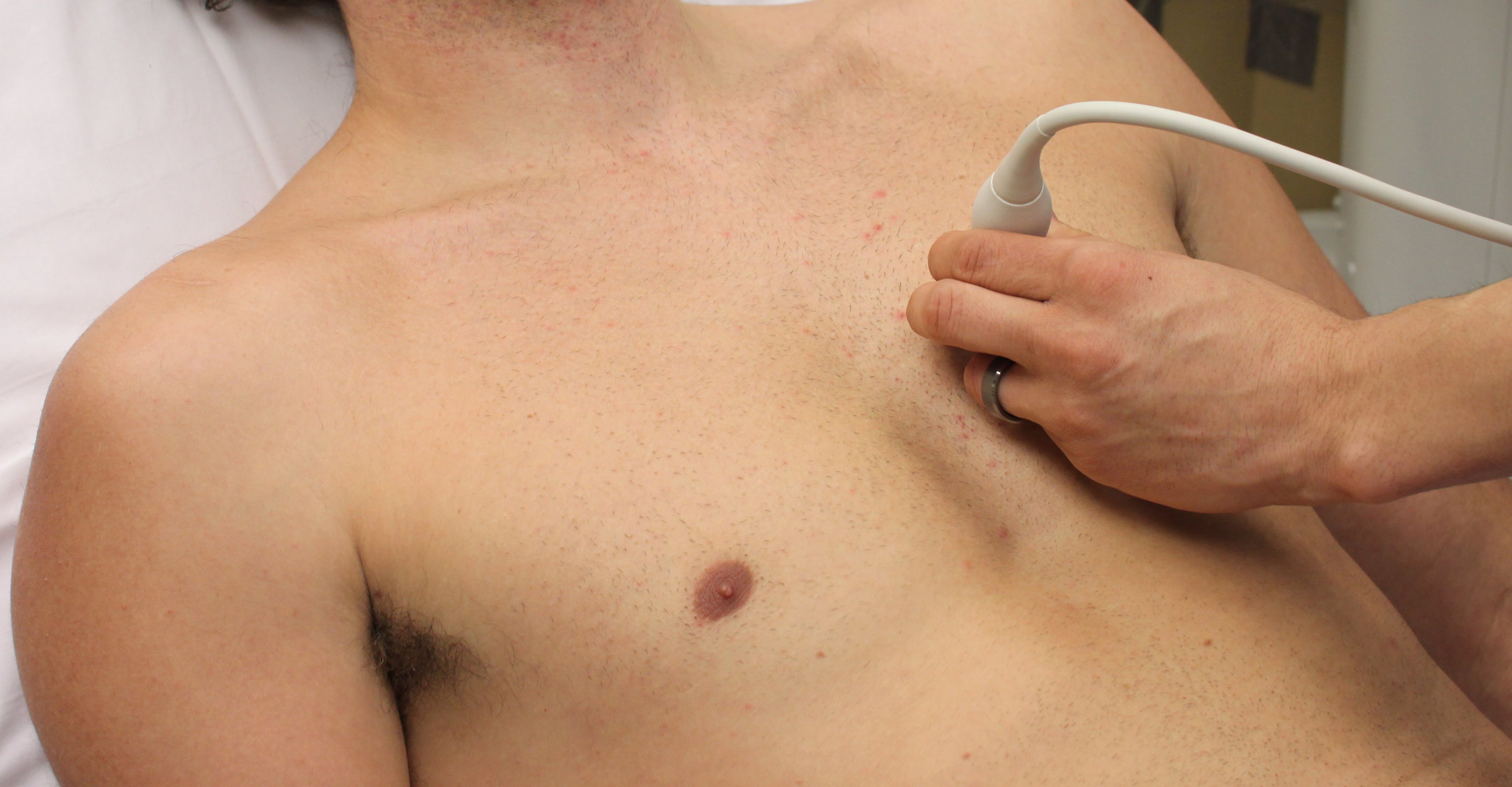
A phased array or curvilinear probe should be used, and each of the eight zones of the lungs should be evaluated including four anterior and four postero-lateral zones (Figure 2,3).
Identifying B-lines is not a difficult examination. One study found that agreement of the presence of b-lines between experienced and novice ultrasonographers had a kappa value of 0.92 (for reference, a kappa value above 0.81 is considered “very good”) [23]. A “positive” scan for pulmonary edema consists of two or more regions of the lung bilaterally with three or more B-lines [22]. The best way to accurately measure the amount of lines present in an area is to freeze the image and cine back to the frame with the most B-lines [24].
Lichtenstein compared auscultation, CXR and lung ultrasound (LUS), and found their accuracies to be 55%, 72% and 95% for interstitial edema. Xirouchaki and colleagues [19] compared US to CXR and found CXR and LUS to have an accuracy for diagnosis of pulmonary edema of 58% and 94%, respectively. These two previous studies used a CT scan as their gold standard, while the next two studies actually used clinical course in the hospital and discharge diagnosis of CHF or not CHF as their gold standard. Liteplo and colleagues [18] found a +LR of infinity and a –LR of 0.78 when all eight lung fields were positive. Prosen and colleagues [17] performed ultrasound on 218 patients that had diagnostic uncertainty between CHF or COPD and found B-lines on LUS to have a +LR of 20. Their lower +LR and better –LR was likely due to the fact that they only required 2 positive lung fields bilaterally before they called it CHF. A recent meta-analysis evaluating the utility of ultrasound for the diagnosis of cardiogenic pulmonary edema found the presence of b-lines to have an impressive sensitivity of 94.1%, a specificity of 92.4%, a +LR of 12 and a –LR of 0.06 [25].
The presence or absence of b-lines bilaterally is great at ruling in or ruling out CHF, but their absence does not necessarily mean the patient has COPD as the cause of their dyspnea. For instance, a patient can have a pneumothorax, pneumonia, anemia, or another diagnosis. If the main diagnostic dilemma is the differentiation of CHF versus COPD, however, the absence of b-lines can push you towards a diagnosis of COPD.
Conclusion:
Differentiating COPD and CHF in an acutely dyspneic patient is an important task that must be done quickly and often with minimal time and minimal resources. Unfortunately, we often make the wrong initial choice. A study by Collins and colleagues [26] found that in a sample of 173 patients that were subsequently diagnosed with heart failure, 33% were misdiagnosed in the ED. The most common factors associated with missed acute decompensated heart failure are a previous history of COPD, no previous history of CHF, and a BNP below 500 [24]. Making the wrong initial diagnosis can be detrimental to these patients, as giving beta agonists to patients with CHF exacerbations has been shown to lead to adverse outcomes, including death [3].
Ultrasound is a fast bedside test which some studies show has immense diagnostic utility. Even with the great +LR of LUS, like any other test or physical exam maneuver we have in medicine, LUS should not be performed and interpreted in a vacuum. It should be used in conjunction with the rest of your history, physical exam and tests. B-lines also are present in a multitude of other pulmonary pathologies including pulmonary contusion, pulmonary infarction, pneumonia, pneumonitis, atelectasis, pulmonary fibrosis, and ARDS.
[bg_faq_start]References:
- Epler GR, Carrington CB, Gaensler EA. Crackles (rales) in the interstitial pulmonary diseases. Chest. 1978;73(3):333-9.
- Piirilä P, Sovijärvi AR, Kaisla T, Rajala HM, Katila T. Crackles in patients with fibrosing alveolitis, bronchiectasis, COPD, and heart failure. Chest. 1991;99(5):1076
- Zeng Q, Jiang S. Update in diagnosis and therapy of coexistent chronic obstructive pulmonary disease and chronic heart failure. J Thorac Dis. 2012;4(3):310-5.
- Oshaug K, Halvorsen PA, Melbye H. Should chest examination be reinstated in the early diagnosis of chronic obstructive pulmonary disease?. Int J Chron Obstruct Pulmon Dis. 2013;8:369-77.
- Mccullough PA, Hollander JE, Nowak RM, et al. Uncovering heart failure in patients with a history of pulmonary disease: rationale for the early use of B-type natriuretic peptide in the emergency department. Acad Emerg Med. 2003;10(3):198-204.
- Jorge S, Becquemin MH, Delerme S, et al. Cardiac asthma in elderly patients: incidence, clinical presentation and outcome. BMC Cardiovasc Disord. 2007;7:16.
- Kataoka H, Matsuno O. Age-related pulmonary crackles (rales) in asymptomatic cardiovascular patients. Ann Fam Med. 2008;6(3):239-45.
- Wang CS, Fitzgerald JM, Schulzer M, Mak E, Ayas NT. Does this dyspneic patient in the emergency department have congestive heart failure?. JAMA. 2005;294(15):1944-56.
- Holleman DR, Simel DL. Does the clinical examination predict airflow limitation?. JAMA. 1995;273(4):313-9.
- Straus SE, Mcalister FA, Sackett DL, Deeks JJ. Accuracy of history, wheezing, and forced expiratory time in the diagnosis of chronic obstructive pulmonary disease. J Gen Intern Med. 2002;17(9):684-8.
- Alrajab S, Youssef AM, Akkus NI, Caldito G. Pleural ultrasonography versus chest radiography for the diagnosis of pneumothorax: Review of the literature and meta-analysis. Crit Care 2013;17:R208.
- Collins SP, Lindsell CJ, Storrow AB, Abraham WT. Prevalence of negative chest radiography results in the emergency department patient with decompensated heart failure. Ann Emerg Med. 2006;47(1):13-8.
- Müller NL, Coxson H. Chronic obstructive pulmonary disease. 4: imaging the lungs in patients with chronic obstructive pulmonary disease. Thorax. 2002;57(11):982-5
- Lichtenstein D, Goldstein I, Mourgeon E, Cluzel P, Grenier P, Rouby JJ. Comparative diagnostic performances of auscultation, chest radiography, and lung ultrasonography in acute respiratory distress syndrome. Anesthesiology. 2004;100(1):9-15.
- Agricola E, Bove T, Oppizzi M, et al. “Ultrasound comet-tail images”: a marker of pulmonary edema: a comparative study with wedge pressure and extravascular lung water. Chest. 2005;127(5):1690-5.
- Gargani L, Lionetti V, Di cristofano C, Bevilacqua G, Recchia FA, Picano E. Early detection of acute lung injury uncoupled to hypoxemia in pigs using ultrasound lung comets. Crit Care Med. 2007;35(12):2769-74.
- Prosen G, Klemen P, Štrnad M, Grmec S. Combination of lung ultrasound (a comet-tail sign) and N-terminal pro-brain natriuretic peptide in differentiating acute heart failure from chronic obstructive pulmonary disease and asthma as cause of acute dyspnea in prehospital emergency setting. Crit Care. 2011;15(2):R114.
- Liteplo AS, Marill KA, Villen T, et al. Emergency thoracic ultrasound in the differentiation of the etiology of shortness of breath (ETUDES): sonographic B-lines and N-terminal pro-brain-type natriuretic peptide in diagnosing congestive heart failure. Acad Emerg Med. 2009;16(3):201-10
- Xirouchaki N, Magkanas E, Vaporidi K, et al. Lung ultrasound in critically ill patients: comparison with bedside chest radiography. Intensive Care Med. 2011;37(9):1488-93.
- Lobo V, Weingrow D, Perera P, Williams SR, Gharahbaghian L. Thoracic ultrasonography. Crit Care Clin. 2014;30(1):93-117, v-vi.
- Silva S, Biendel C, Ruiz J, et al. Usefulness of cardiothoracic chest ultrasound in the management of acute respiratory failure in critical care practice. Chest. 2013;144(3):859-65.
- Volpicelli G, Elbarbary M, Blaivas M, et al. International evidence-based recommendations for point-of-care lung ultrasound. Intensive Care Med. 2012;38(4):577-91.
- Cibinel GA, Casoli G, Elia F, et al. Diagnostic accuracy and reproducibility of pleural and lung ultrasound in discriminating cardiogenic causes of acute dyspnea in the emergency department. Intern Emerg Med. 2012;7(1):65-70
- Anderson KL, Fields JM, Panebianco NL, Jenq KY, Marin J, Dean AJ. Inter-rater reliability of quantifying pleural B-lines using multiple counting methods. J Ultrasound Med. 2013;32(1):115-20.
- Al deeb M, Barbic S, Featherstone R, Dankoff J, Barbic D. Point-of-care Ultrasonography for the Diagnosis of Acute Cardiogenic Pulmonary Edema in Patients Presenting With Acute Dyspnea: A Systematic Review and Meta-analysis. Acad Emerg Med. 2014;21(8):843-852.
- Collins SP, Lindsell CJ, Peacock WF, Eckert DC, Askew J, Storrow AB. Clinical characteristics of emergency department heart failure patients initially diagnosed as non-heart failure. BMC Emerg Med. 2006;6:11.
Reviewing with the Staff | Reviewed by Dr. Daniel Kim MD FRCPC
Dr. Kim is the Ultrasound Fellowship Director, University of British Columbia and an Emergency Physician, Vancouver General Hospital
He is a graduate of the University of Toronto’s Royal College emergency medicine residency program. He completed an ultrasound fellowship at Denver Health Medical Center. Dan is currently an emergency physician at Vancouver General Hospital as well as the ultrasound fellowship director at the University of British Columbia. It goes without saying that his academic interest is… all things ultrasound!
Nice write up, and great case illustrating a conundrum that every emergency physician has experienced: does this dyspneic patient have COPD or CHF? In this scenario, it’s not unusual for a patient to receive antibiotics, steroids, albuterol, furosemide, and nitroglycerin – just to “cover all the bases.” But are we doing good for the patient?
As Jacob reminds us, the accuracy of our physical exam for diagnosing lung pathology is mediocre. CHF patients may have wheezing instead of crackles, and the opposite is true of some COPD patients. However, we should remember that our individual physical exam tests and findings do not occur in a vacuum. Instead, they are one piece of the puzzle that needs to be combined with a good history and appropriate testing for us to come to the right final diagnosis. In fact, Wang’s JAMA systematic review found that a high pretest probability for heart failure (based on clinician gestalt) had a positive LR of 9.9 for a final diagnosis of heart failure. We should feel reassured that our clinical judgment is valuable! But we aren’t perfect. An intermediate or low initial clinical suspicion decreased the likelihood of heart failure (LR 0.65) but did not exclude it. <1>
The chest x-ray is also an imperfect test, given that up to 1 in 5 patients admitted from the ED with acute decompensated heart failure have no signs of congestion on x-ray. <2> So where does this leave us?
Ultrasound provides us with a rapid, noninvasive, and radiation-free way of ruling in or ruling out pulmonary edema at the bedside. But is it accurate? Al Deeb’s recent systematic review indicates that it is. The pooled sensitivity and specificity of B-lines for pulmonary edema is 94% and 92% respectively. This translates to a positive LR of 12.4 and a negative LR of 0.06. <3> This can help to significantly change the pretest probability of disease to a posttest probability that gives us confidence in our diagnosis. While there are some issues with Al Deeb’s meta-analysis (like heterogeneity), there’s no doubt in my mind that ultrasound is accurate. Laursen showed that an ultrasound protocol for dyspnea provides the correct diagnosis earlier than usual diagnostic testing (88% in the ultrasound group vs 64% in the control group had the correct diagnosis at 4 hours after admission). There was no difference in mortality, but his study wasn’t powered for this endpoint. <4> Hopefully, future research is able to demonstrate improvements in patient oriented outcomes (like mortality). In the interim, we should use all the tools at our disposal to come up with the right diagnosis to provide the right treatment – and avoid unnecessary (and potentially harmful) treatment that “covers all the bases.”
References
- Wang CS, FitzGerald JM, Schulzer M, et al. Does this dyspneic patient in the emergency department have congestive heart failure? JAMA 2005; 294:1944-56.
- Collins SP, Lindsell CJ, Storrow AB, et al. Prevalence of negative chest radiography results in the emergency department patient with decompensated heart failure. Ann Emerg Med 2006; 47:13-8.
- Al Deeb M, Barbic S, Featherstone R, et al. Point-of-care ultrasonography for the diagnosis of acute cardiogenic pulmonary edema in patients presenting with acute dyspnea: a systematic review and meta-analysis. Acad Emerg Med 2014; 21:843-852.
- Laursen CB, Sloth E, Lassen AT, et al. Point-of-care ultrasonography in patients admitted with respiratory symptoms: a single-blind, randomised controlled trial. Lancet Respir Med 2014; 2:638-46.


