
You may be wondering; why do we need another post on left ventricular assist devices ( LVADs ) when there are already several phenomenal articles out there in the #FOAMed universe? There are two reasons for this. First, to my knowledge, there has not yet been a #FOAMed article regarding community emergency department (ED) management of LVAD related complications. LVADs are surgically placed in tertiary care, academic centres with specialized LVAD teams. However, the patients who receive LVADs are not confined to those centres.1 Second, in this article, we have expert contribution from Dr. Gil Kimel, a palliative care physician with fellowship training in palliative management of congestive heart failure, as well as Annemarie Kaan, a masters-prepared nurse, who works as part of the LVAD team at St. Paul’s Hospital in Vancouver.
The purpose of this post is to provide a practical, case based approach to the patient with a LVAD who presents to the ED. This is not intended to be comprehensive as there are already several detailed articles out there [see below].
Brief Background on LVADs
Fully implantable LVADs are mechanical devices that provide circulatory support in patients with end stage left sided heart failure. They are generally placed for three reasons1–3:
- Bridge to recovery
- In British Columbia (BC), about 10% are able to be explanted with cardiac recovery (Annemarie Kaan, Oct 27, 2016).4
- Bridge to transplantation (most common)
- Destination therapy (e.g. not suitable for transplant but refractory to medical management)
- In BC this does occur occasionally in the setting of a patient who initially was a transplant candidate but who became ineligible during the period of time after LVAD implantation(Annemarie Kaan, Oct 27, 2016).4
First generation devices provided pulsatile flow with a pump mechanism. Newer, second generation devices, provide continuous flow, usually with a centrifugal or axial flow generator.2 Currently, patients with LVADs have second generation devices.2 In BC, the HeartWare HVAD device is used (Annemarie Kaan, Oct 27, 2016); the HeartMate II device is the other main implantable device used in Canada.3,4
At the present time, approximately 160 adult heart transplants occur per year in Canada, and of those, about 28 are performed in BC.4,5 In contrast, there are approximately 90,000 patients in BC living with heart failure.6
Generally speaking, there are five components to an LVAD [Figure 1]:
- The pump
- The inflow cannula
- The outflow cannula
- The driveline
- The controller and power source

Figure 1: HeartWare HVAD device in-situ.7
The inflow cannula is connected to the left ventricular apex, the pump creates forward flow, the outflow cannula is connected to the ascending aorta, and the driveline exits the body and connects to the controller which is connected to the power source.2
In the device shown above, there is a centrifugal pump powered by an impeller that creates continuous flow [Figure 2].
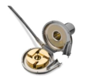
Figure 2: HeartWare HVAD pump showing impeller.7
Chronic anticoagulation, with warfarin, and anti-platelet therapy, usually aspirin, is required for patients with LVADs in situ.2 Target INR is generally 2-2.5 and, as such, bleeding complications can occur.2,4
[bg_faq_start]Case 1: The vital signs
You are the emergency physician on duty in a community ED when the triage nurse approaches you in a panic stating, “I have a patient who I cannot seem to get a blood pressure, oxygen saturation, or a pulse on but they are talking to me. Can you come right away?”
LVADs are inserted in patients with end stage left heart failure and therefore, depending on their residual function, they may not have a palpable pulse, blood pressure, or oxygen saturation by pulse oximeter.2,3
If you are unable to obtain a mean arterial pressure (MAP) using an electronic NIBP device, you should use a manual blood pressure cuff and a Doppler flow meter.2 Using either the brachial or radial artery with the Doppler, inflate the cuff until the sound of blood flow is extinguished. Then, release the cuff until the sound returns. The point at which the sounds returns approximates the mean arterial pressure. This should be between 70-80 mmHg.2
Pulse oximetry is often unattainable and is considered unreliable if the pulse is subtle as is the case with many LVAD patients.2 It is possible to use cerebral oximetry3 to obtain oxygen saturation or, if needed, you can pursue arterial blood gas or even arterial line placement for the most accurate oxygenation and blood pressure measurements.2
[bg_faq_end][bg_faq_start]Case 2: The MAP points to the sky
One week after case one, you are surprised to see another LVAD patient in your ED who complains of generalized headache. Her vital signs, measured as described above, are within normal limits except for the MAP which is 105 mmHg. You wonder, at what point is the MAP too high and what do I need to do?
Hypertension, in the context of an LVAD, is defined as a MAP of greater than 90 mmHg.2 This portends a higher risk of stroke and declining function of the device.2
If the MAP is >90 mmHg, it should be treated with afterload reduction such as angiotensin converting enzyme inhibitors. 2Depending on the clinical context, this may need to be done urgently with intravenous agents. I would recommend contacting your local VAD centre prior to initiating therapy. In BC, the VAD Hotline can be reached 24/7 at 604-250-2658.
[bg_faq_end][bg_faq_start]Case 3: Hypotension
Your unfortunate patient from Case 2 now complains of feeling very light headed. You re-check the vital signs and notice the MAP is 45 mmHg. Your main concern is that you may have over treated the hypertension, but you begin to think of other causes and order a work-up. Your ECG shows the following [Figure 3].

Figure 3: ECG concerning for ventricular tachycardia
You wonder two things. Does dysrhythmia matter when the patient has an LVAD supporting their circulation? What LVAD specific complications can cause hypotension (defined as MAP < 60 mmHg)?1
Tachydysrhythmias are not uncommon in patients with LVADs and occur 22-59% of the time.3 Because of this, most LVAD patients will have an implantable cardioverter/defibrillator device inserted at the time of LVAD implantation.3 However, if you happen to see a patient as described above, you need to treat the dysrhythmia. Prolonged tachydysrhythmias in LVAD patients can lead to right ventricular failure with a subsequent decrease in left ventricular preload and hypotension.2 Therefore, symptomatic ventricular tachycardia or ventricular fibrillation should be treated urgently [2,3]. Again, I would suggest contacting your VAD centre prior to initiating therapy. If unstable, electrical cardioversion should be performed immediately.2
There is a myriad of problems that can cause hypotension in patients with LVADs. I am not going to cover all of them in this article. However, here is a list of potential causes for which the articles listed at the bottom of this article are an excellent resource:
- Dehydration
- Tamponade (mostly seen early after implantation)
- Right sided heart failure
- Arrhythmias
- Bleeding
- Infection
- Thrombosis
- Device failure
The ones I have highlighted have potential interventions that you can get started in a community setting in conjunction with your nearest LVAD centre and we will go through these briefly. The others, we have already covered.
Thrombosis
- Suspect this diagnosis in anyone with an LVAD and MAP < 60mmHg1
- Thrombosis occurs in <10% of LVAD patients in BC5
- Add hemolytic work-up (lactate dehydrogenase, urinalysis, plasma-free Hgb) if suspecting thrombosis as these often show a hemolytic pattern2
- Echocardiography will occasionally be able to pick this up2
- In conjunction with your nearest LVAD centre, consider starting anticoagulation with intravenous heparin and potentially thrombolysis in conjuction with your local VAD centre2
Bleeding
- Bleeding most commonly occurs in the gastrointestinal tract3
- Work up as usual paying particular attention to coagulation parameters
- Consult your nearest VAD centre as well as gastroenterology to guide further management with regard to reversing coagulopathy
Infection
- Infection relating specifically to the LVAD is common2
- It can involve any part from the driveline, to the pocket where the pump is housed, to the cannulae
- Suspect infection whenever an LVAD patient presents with fever, pain at driveline exit site, general malaise, or abdominal pain [2]
- Consult your nearest LVAD centre promptly as surgical management may be necessary
- Consider CT or ultrasound to look for device related abscess2
- Cover for Gram positive and Gram negative organisms after drawing blood cultures2,4
- Consider coverage for fungal organisms as they make up 9% of LVAD infections and carry the highest mortality2
Device failure
- The most important step here is to check the controller for any alarms and for battery power [Figure 4]
- The second most important step is to immediately contact your nearest LVAD centre and the patient’s caregiver
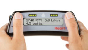
Figure 4: HeartWare HVAD control box.7
Tamponade
- Consider this in any hypotensive LVAD patient presenting shortly after device implantation
Case 4: Cardiac arrest
The day really ends poorly for you and your patient as you get called into the resuscitation bay because your patient has become unconscious. You re-check the vital signs and cannot obtain a MAP. You auscultate for the “hum” of the LVAD and hear nothing. You perform a bedside ultrasound which reveals no evidence of a “suck down” event. You’ve heard that chest compressions are contraindicated in LVAD patients and wonder what to do next?
The important factors here are to check the control box for power and to auscultate the chest.2 If there is no power, then connect the device to a battery or power station if possible. If there is no “hum”, the device is not functioning.2
If there is no “hum”, the power has been checked, and there is a MAP of zero, then start chest compressions! There is a risk of dislodging the cannulae with the trauma of chest compressions but this is not a contraindication in the above scenario.2,3 After initiating chest compressions, follow ACLS protocols as usual including ruling out reversible causes. If you are running a cardiac arrest in an LVAD patient and you have a cardiovascular surgery service in your hospital, contact them stat!
[bg_faq_end]Putting it all together
If you encounter a patient in your ED who has an LVAD and you suspect a complication:
- Ask the patient or their family for the info card for their device as well as the contact details for their VAD specialist and contact them (In BC, the 24/7 VAD Hotline can be reached at 604-250-2658).
- If unable to obtain blood pressure by NIBP, use Doppler with a manual cuff.
- If pulse oximetry is not reliable, then pursue cerebral oximetry, ABG, or an arterial line depending on the scenario.
- If hypotensive, obtain a work-up including basic CBC, electrolytes, coagulation panel, chest xray, and ECG. Consider adding hemolytic work up and performing bedside ultrasound. Administer an IV fluid bolus or PO fluids if awake and alert.
- If you suspect infection, draw blood cultures, cover with broad spectrum antibiotics, and resuscitate as appropriate.
- Sustained symptomatic ventricular tachycardias/ventricular fibrillation should be treated urgently with cardioversion. Defibrillator pads should not be placed directly over the LVAD device.
- If there is no “hum” on chest auscultation and/or there are no signs of perfusion, begin chest compressions and contact a cardiac surgeon if one is available on site.
There are many excellent resources out there if you would like to pursue more in depth information about this topic; please see the list below. As well, Dr. Rahul Patwari has put together two excellent YouTube videos for the visual learners out there. I highly recommend them as they are succinct, illustrated, and easy to understand (LVAD 1 & LVAD 2).
Additional Resources on LVADs
Here are some additional readings that might provide you with more relevant information:
- Here is a nice concise summary of LVAD emergencies by Dr. Zack Shinar on EMCrit from 2012.
- RebelEM put out an excellent article in 2015 going into a bit more detail on the statistics and background of LVADs as well as the ins and outs of complications.
- In this EPMonthly article Zack Shinar gives a scenario based overview of LVADs and their complications.
- Here is a case based 2015 article from Dr. Sumintra Wood on EMDocs discussing LVADs in the ED.
- Chris Nickson authored a 2015 piece on Life In The Fast Lane with all the nitty gritty details of the physics and engineering behind LVAD devices and their complications.
- EM Nerd (Dr. Rory Spiegel) published an article on EMCrit in 2013 that will satisfy even the largest clinical epidemiology and EBM appetite on LVADs.
This post was copyedited by Michael Bravo (@bravbro).
References
LVAD management for EM physicians (by Dr. Kobulnik)
As pointed out by Jared, patients with left ventricular assist devices have unique physiology. As contemporary devices spin blood continuously, the majority of patients with a left ventricular assist device do not have a palpable pulse.1 This leads to some practical issues. Automated and manual blood pressure cuffs often do not yield accurate measurements and tachycardia or bradycardia can really only be detected with an ECG. Moreover, since the difference between systolic and diastolic blood pressure is very small in these patients, the blood pressure measured by Doppler essentially represents systolic, diastolic and mean arterial blood pressure. Importantly, while a Doppler derived pressure of 70mmHg is concerning in patients without an LVAD, that would be a perfectly reasonable (and probably ideal) blood pressure in a patient without a pulse who was supported with a LVAD.1,2 Conversely, a Doppler derived blood pressure of 110mmHg in such a patient is dangerously high and should prompt immediate medical attention.2 The absence of a palpable pulse does come with an upside. As there is seldom a visible carotid pulsation, the jugular venous pressure becomes much more easily discernable.
Generally, patients with LVADs ought to be treated much the same way that other patients with advanced heart failure are treated. Chronically, they are often managed with ACE-inhibitors, beta-blockers, aldosterone antagonists, anti-coagulants and antiplatelet therapy, as well as Implantable Cardioverter Defibrillators (ICDs) and cardiac re-synchronization therapy (CRT).2 Any patient with an LVAD presenting to the emergency room should be evaluated for the usual things that can affect a patient with heart failure in addition to specific complications associated with the device. Specifically, they should be assessed for:
- uncompensated heart failure,
- a concomitant infection,
- adverse effects of their medications including volume depletion/hypotension, bradycardia, renal dysfunction, associated electrolytes disturbances and anemia.
As patients with LVAD’s are prone to blood born infections and may not have a conspicuous sepsis syndrome because of unknown reasons1 there should be a very low threshold for obtaining blood cultures. If there is a clinical suspicion of infection, such as fever, general malaise, or purulent drainage from the driveline site, empiric antibiotics can be initiated, after blood cultures are obtained, of course.1
Finally, while anemia is common in patients with heart failure, related to chronic disease and anti-coagulation,3 patients with left ventricular assist devices are particularly prone to anemia both because of associated hemolysis and the consumption of large Von Willebrand multimers.4,5
Patients presenting with uncompensated heart failure should be given diuretics. Patients supported with an LVAD tend to tolerate ventricular arrhythmias well, but as the right ventricle is not supported, these should be treated, and generally can be treated similarly to patients that are unsupported.2 Patients with symptomatic anemia should be transfused, and may require concomitant diuresis. Adjustment of their anti-coagulation and anti-platelet therapy may be necessary but should be done in consultation with an LVAD center, as the risk of thrombosis needs to be integrated into the decision-making.
Mechanical failure of contemporary devices is rare. Diagnosis requires both the integration of clinical findings, device parameters/alarms, laboratory data and along with an echocardiogram. In an unwell but hemodynamically stable patient, the evaluation for this is urgent but not emergent and should be done in consultation with a local LVAD center. In a hemodynamically unstable patient, especially if the device itself is alarming or when a VAD hum cannot be heard, a change in power source and/or a controller swap are appropriate initial steps.1 The patient’s caregiver can often assist with this. As a mechanical failure of the device or device thrombosis may require urgent surgical intervention, cardiac surgery should be consulted immediately in any hemodynamically unstable patient with an LVAD device.1
It is important to recognize that LVADs can be protective and some patients with the devices tolerate medical illness better than many unsupported patients with heart failure. Moreover, as the patients and their caregivers go through intense teaching prior to discharge,2 they are usually quite informed and may be helpful in their own assessment in the emergency room.
1. Robertson J, Long B, Koyfman A. The emergency management of ventricular assist devices. Am J Emerg Med. 2016;34(7):1294-1301.
2. Feldman D, Pamboukian SV, Teuteberg JJ, et al. The 2013 International Society for Heart and Lung Transplantation Guidelines for mechanical circulatory support: executive summary. J Heart Lung Transplant. 2013;32(2):157-187.
3. Tim Goodnough L, Comin-Colet J, Leal-Noval S, et al. Management of anemia in patients with congestive heart failure. Am J Hematol. 2017;92(1):88-93.
4. Sajgalik P, Grupper A, Edwards BS, et al. Current Status of Left Ventricular Assist Device Therapy. Mayo Clin Proc. 2016;91(7):927-940.
5. Patel CB, Cowger JA, Zuckermann A. A contemporary review of mechanical circulatory support. J Heart Lung Transplant. 2014;33(7):667-674.


Jared Baylis
Latest posts by Jared Baylis (see all)
- First Year Diaries E04: Dr. Daniel Ting & Dr. Jared Baylis – Transitioning to EM Practice - June 22, 2020
- Learning to fly in #PGWHY5: Five tips for survival in the exam year - July 19, 2019
- The Canadian Enhanced Competency Directory - August 4, 2017