This post has been co-written by Drs. Kathy Boutis and Maxim Ben Yakov.
The dogmas of the past are now being challenged for the most common minor pediatric fractures, distal radius buckle fractures and minor distal fibular fractures. Since these fractures are stable and have an excellent prognosis, they do not need to be routinely immobilized in a cast nor followed by an orthopedic surgeon. Last time, we reviewed low risk distal radius fractures in kids (read that piece here). This article reviews the evidence that recommends that management of the low risk pediatric ankle fractures.
Low Risk Pediatric Ankle Fractures
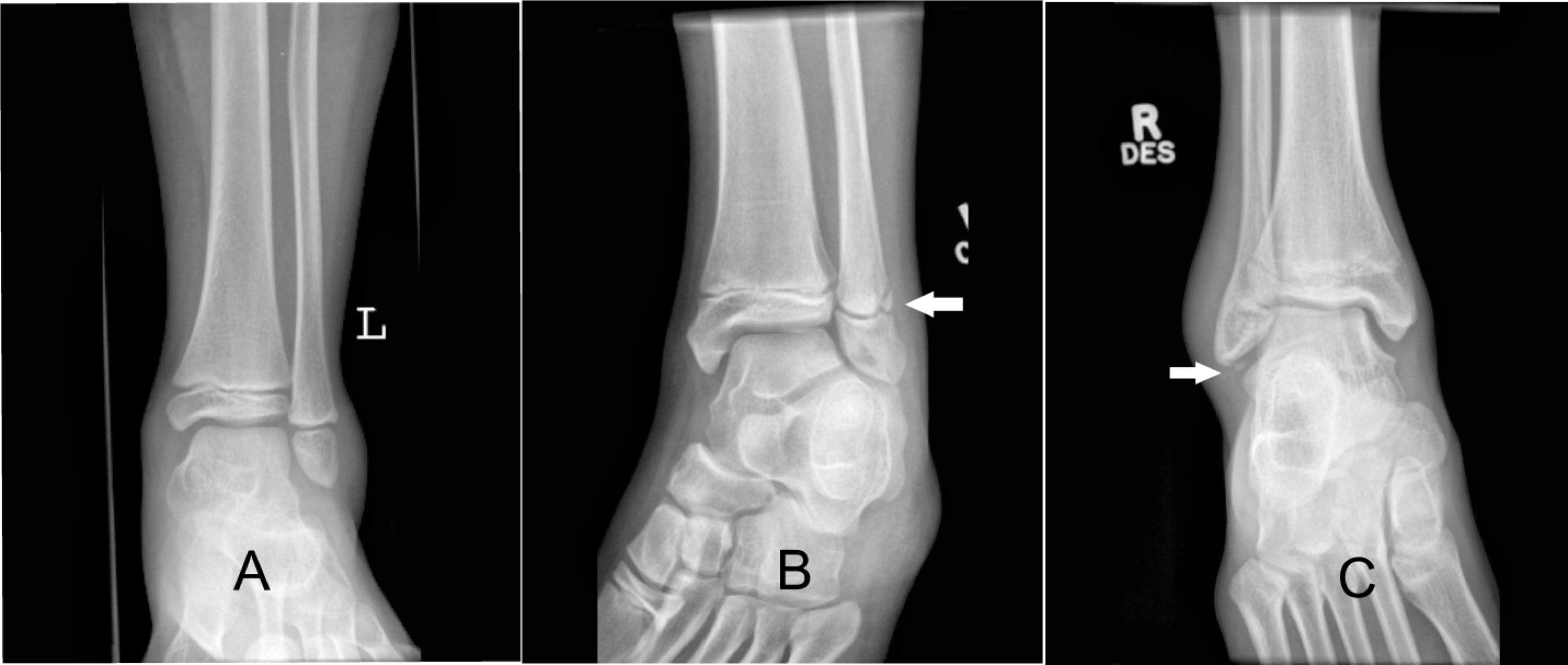
Low risk ankle fractures are the most common lower-extremity fractures in children, and include isolated undisplaced distal fibular Salter-Harris I, II or avulsion fractures.1,2 They have excellent functional outcomes and are exceptionally low risk for future complications.

Figure 1: Low Risk Ankle Fractures (A) Presumed distal fibular Salter-Harris I physeal fracture (B) Distal fibular Salter-Harris II physeal fracture (C) Distal fibular avulsion fracture
There are three randomized control trials which demonstrate that management of these low-risk ankle fractures with a removable device (ankle brace or ace wrap) and a self-regulated return to activities was superior to a fiberglass cast/back-slab for three weeks with respect to recovery.2–4 There was also a parental preference for removable devices and treating patients using an ankle brace versus a cast was found to be cost-effective for the health care system.2 Finally, similar to distal radius buckle fractures, follow up with an orthopedic surgeon is not routinely necessary for these injuries and should be reserved for cases that are not improving as expected.5,6
It is also important to note that of these low risk fractures, the distal fibular Salter-Harris I fracture is a lot rarer than previously thought. Due to the presumed weakness of the growth plate relative to adjacent ligaments, skeletally immature children with isolated swelling and tenderness over the distal fibula and no radiographic evidence of fracture are often diagnosed with Salter-Harris I fractures. Once this diagnosis is made clinically, traditional management has included casting and follow-up by an orthopedic surgeon. However, a study that included 135 children with a clinical presentation consistent with a distal fibular Salter-Harris I fracture had MRI imaging of the injured ankle and results demonstrated that in fact only 4 (3.0%) of these children had Salter-Harris I fractures, whereby 2 were only partial injuries.7 Rather, all children had sprain injuries, including the ones with Salter-Harris I fractures, and about 67% of the sprain injuries were moderate to high-grade sprain injuries and occasionally associated with radiographically occult distal fibular fractures. But, regardless of the specific MRI diagnosis, all children recovered well when treated with a removable ankle brace and self-regulated return to activities.7 This adds to the evidence derived by the randomized control trials that these injuries are safely managed with minimal interventions and follow up with the primary care physician.


