
An elbow fracture is a common pediatric injury in emergency medicine. These fractures present challenges in diagnosis due to the maturing skeletal anatomy and the subtlety of these injuries.1 Plain radiographs are adequate to detect elbow fractures in most cases and a systematic approach can help learners diagnose these pathologies.2,3
An approach to reading elbow radiographs:
After confirming patient identification and appropriate radiographic view, a mnemonic to help guide your approach to elbow fractures is to ask yourself, “is this a CASE of an elbow fracture”?
C– CRITOE: Are the 6 ossification centers of the elbow joint normal?
A–Alignment: Assess for fractures and/or dislocations.
S– Subtle Fractures: Ensure no subtle fractures have been overlooked.
E– Effusions: Is there evidence of a joint effusion?
C– CRITOE.
Are the 6 ossification centers of the elbow joint normal? These include, the Capitellum, Radius, Internal (medial) epicondyle, Trochlea, Olecranon, and External (lateral) epicondyle (refer to figure 1). The ages at which these centers appear and fuse vary with age.2,3

Figure 1. Ossification centers. Photo Credit: The Radiology Assistant1
A–Alignment.
Assess for fractures and/or dislocations. This can be investigated by looking at the alignment of the:
(1) anterior humeral line (refer to figure 2) and,
(2) the radiocapitellar line (refer to figure 3).

Figure 2. The anterior humeral line. A vertical line from the anterior surface of the humerus should divide the middle third of the capitellum. Photo Credit: The Radiology Assistant1

Figure 3. The radiocapitellar line. A horizontal line should pass through the centers of the capitellum and radial neck. Photo Credit: The Radiology Assistant1
S– Subtle Fractures.
Ensure no subtle fractures have been overlooked (refer to figure 4).1 Supracondylar fractures account for up to 60% of pediatric elbow fractures. The epidemiology of other injuries varies and include lateral epicondyle fractures (10-20%), proximal radial and olecranon fractures (5-8%) and medial epicondyle fractures (<1-10%).1–4
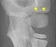
Figure 4. An example of a subtle lateral condyle fracture. Photo Credit: The Radiology Assistant1
E– Effusions.
Is there evidence of a joint effusion? This can be examined by looking for:
(1) increased anterior lucency and elevation of the anterior fat pad, and/or
(2) manifestation of the posterior fat pad (refer to figure 5).1–3
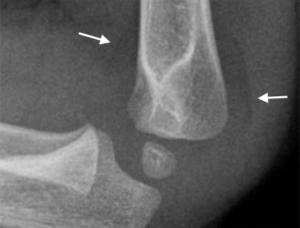
Figure 5. Anterior and posterior fat pad. Photo Credit: The Radiology Assistant1
This post was uploaded by Sean Nugent (@sfnugent)
[bg_faq_start]References
Reviewing with the Staff
Elbow fractures are one of the most common pediatric injuries we see in Emergency Medicine. As noted above, x-ray interpretation of elbows is often challenging for learners, because of evolving anatomy and the subtlety of some elbow fractures (1). In addition to a fundamental understanding of anatomy, bony development and x-ray features of pediatric elbows, having a standardized approach (as above) is advantageous for accurate and timely interpretation. This will also impress your supervising physician!
A few other tips…
• A posterior effusion is always abnormal. One should suspect an occult fracture, if no obvious fracture is seen.
• The most common pediatric elbow fracture is a supracondylar fracture.
• A thorough neurovascular examination is vital to rule out potential injuries to the brachial artery, median, radial and/or ulnar nerves.

Graham Wilson
Latest posts by Graham Wilson (see all)
- Tiny Tip: Is this a ‘CASE’ of an elbow fracture? - June 23, 2017