
Welcome back to PoCUS Previews, your illustrated guide to the world of Point of Care Ultrasound (PoCUS)!
In the hands of a skilled sonographer, PoCUS can serve as a valuable tool while assessing a patient in trauma. PoCUS can quickly and fairly accurately detect blood loss and signal the presence of internal organ damage. Thus, the Focused Assessment with Sonography in Trauma (FAST) scan is one of the most celebrated uses of PoCUS in the ED. The latest issue of PoCUS Previews gives you a brief intro to just that!
Background and Indications
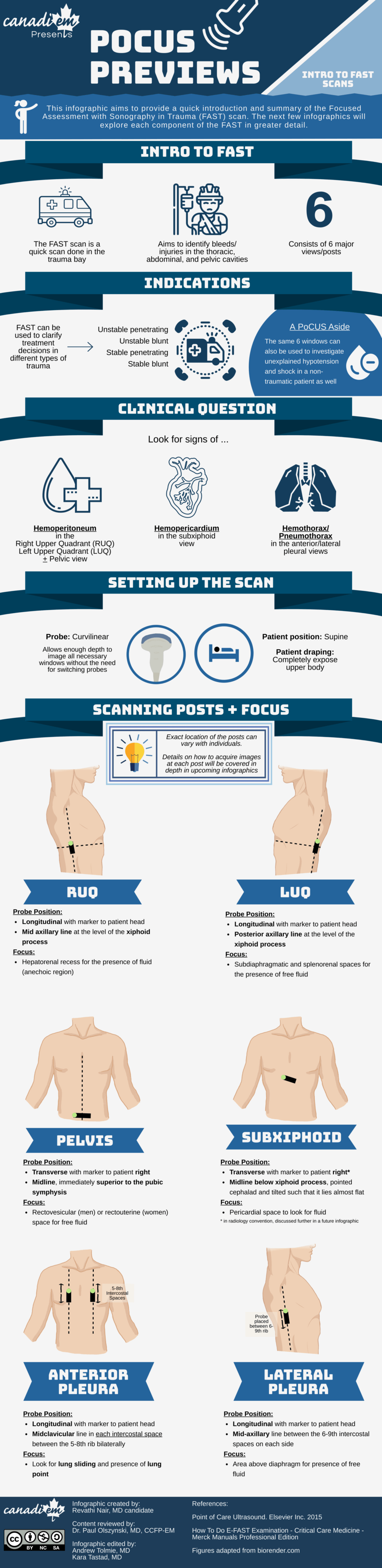
The FAST scan is done to work up patients presenting with stable and unstable penetrating or blunt traumas. Historically, it served to look for signs of internal bleeding in 2 different areas: the peritoneum, and the pericardium.
The bleeding in the peritoneum is assessed using 3 peritoneal windows: the right upper quadrant (RUQ), left upper quadrant (LUQ) and the pelvic window. The presence of hemopericardium (or blood in the pericardium) is assessed using a subxiphoid view of the heart.
Over time, FAST scans started including views of the lateral lungs to assess for hemothorax and anterior lungs for assessment of pneumothorax. Initially, the addition of these views to the FAST scan was known as extended FAST (or e-FAST). However, with time these views became so commonplace that they were assumed to be a part of any regular FAST scan you receive. Hence, even though the lung views are included in the infographic, we have still decided to refer to all 6 windows as the FAST scan.
Setting up a FAST Scan
The entire scan is done with the patient in a supine position using the curvilinear probe. Using the curvilinear probe allows one to sufficiently probe the deeper cavities such as the abdomen without the need for switching probes during the assessment, saving us valuable time. This has some important connotations for the cardiac view which we will touch upon further in a future infographic.
We hope this infographic comes in handy the next time you get called into the trauma bay to assess a patient. Stay tuned for in-depth breakdowns of each of the 6 windows of the FAST scan in upcoming PoCUS Previews!
As always, we would love to hear any feedback from you to help improve our future infographics in the series.
Look out for the next infographic in the series: An in-depth breakdown of the RUQ/LUQ windows of the FAST scan!

Bibliography
Soni NJ, Arntfield R, Kory P. Point of Care Ultrasound. Elsevier Inc. 2015
Pace J, Arntfield R. Focused assessment with sonography in trauma: a review of concepts and considerations for anesthesiology. Can J Anesth. 2018 Apr 1;65(4):360–70.
Staff Reviewer
When assessing a patient with blunt or penetrating trauma, it is important to rapidly assess for life-threatening thoracabdominal injuries including pneumothorax, hemothorax, hemopericardium and traumatic pericardial effusion. By incorporating the FAST scan into the initial assessment of the patient, time-to-diagnosis can be shortened, allowing potentially life-saving interventions to be initiated at the earliest opportunity.


Revathi Nair
Latest posts by Revathi Nair (see all)
- CAEP Capsule 23: Day 3 (May 30th-The Finale) - June 3, 2023
- CAEP Capsule 23: Day 2 (May 29th) - May 31, 2023
- CAEP Capsule 23: Day 1 (May 28th) - May 29, 2023