
A 64 year old woman presents to the emergency department with dyspnea. On exam she is mildly tachypneic, has an oxygen saturation of 94% on 2L nasal prongs, and bilateral crackles to auscultation. You suspect acute heart failure and wonder about the role of lung ultrasound as you await her chest x-ray.
Clinical question: What is the sensitivity and specificity of lung PoCUS compared to chest radiograph for diagnosis of acute decompensated heart failure?
Dyspnea is a common ED presentation with a broad differential diagnosis (see quick CanadiEM primer here!). Acute decompensated heart failure is the cause of dyspnea in up to 40% of older adults in the ED.1 Accurate and timely diagnosis of CHF in the ED leads to earlier treatment initiation and disposition for patients, improving quality of care and ED flow.
Previous work has indicated that chest radiography can produce false negative rates of around 20% and waiting for radiographs often results in a delayed diagnosis. A number of studies have evaluated the sensitivity and specificity of chest radiographs for heart failure. Sensitivity ranged between 53-75% while specificity ranged between 86 and 96%.2–5 While there is certainly a role in obtaining radiographs for documentation and confirmation of findings, can ultrasound provide us with accurate and timely information in the ED?
Lung PoCUS
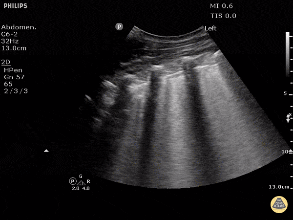
Ultrasound machines are readily available in the ED and operators can produce images more quickly than waiting for radiography. Bilateral lung bases are examined for the presence of b-lines and pleural effusion. The images below show normal lung tissue, B lines and a pleural effusion, as seen on PoCUS.5

Figure 1: A-lines representing normal lung tissue 6

Figure 2: Presence of B Lines 7
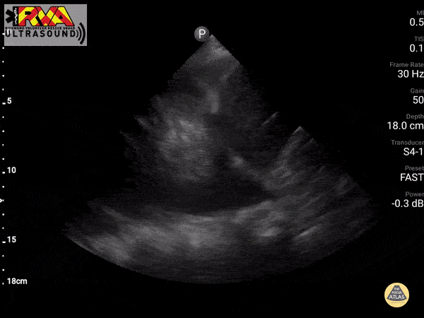
Figure 3: Pleural effusion 8
The Evidence
Two recent papers evaluated the efficacy of lung ultrasound for acute heart failure. Nakao et al. evaluated the accuracy of PoCUS compared to chest x-ray in older adults with dyspnea or cough in a cohort study of 81 patients. They demonstrated a sensitivity of 92.5% (83.4%-97.5%) and specificity of 85.7% (57.2%-98.2%) for lung ultrasound, compared to sensitivity of 63.6% (50.9%-75.1%) and specificity 92.9% (66.1%-99.8%) of chest x-ray in detecting heart failure.4
Additionally, a systematic review and meta-analysis by Maw et al. looked at the accuracy of lung ultrasound compared to chest x-ray for heart failure. Six studies with 1827 total patients were identified. Among patients presenting with dyspnea who had both a lung ultrasound and chest x-ray completed, there was an absolute difference in sensitivity of 15% between PoCUS (Sn 88%, 95%CI 75%-95%) and chest x-ray (Sn 73%, 95%CI 70%-76%).9 There was comparable specificity between PoCUS 90%(95%CI 75%-91%) and chest x-ray 90% (95%CI 75%-97%).9
It is important to recognize that B-lines are non-specific and can also be caused by other pathologies such as pulmonary fibrosis, interstitial pneumonia, and ARDS. Always consider clinical context when interpreting your PoCUS images. Bilateral B-lines are generally more pathognomonic of CHF versus other pathologies.
Bottom Line
Lung ultrasound is more sensitive and similarly specific in comparison to chest x-ray, and can be considered a useful adjunct to clinical exam in the diagnosis of acute heart failure.
Copyedited by Danny Ma (@DannyMa_).
References
- 1.Ray P, Birolleau S, Lefort Y, et al. Acute respiratory failure in the elderly: etiology, emergency diagnosis and prognosis. Crit Care. 2006;10(3):R82. doi:10.1186/cc4926
- 2.Wang C, FitzGerald J, Schulzer M, Mak E, Ayas N. Does this dyspneic patient in the emergency department have congestive heart failure? JAMA. 2005;294(15):1944-1956. doi:10.1001/jama.294.15.1944
- 3.Mueller-Lenke N, Rudez J, Staub D, et al. Use of chest radiography in the emergency diagnosis of acute congestive heart failure. Heart. 2006;92(5):695-696. doi:10.1136/hrt.2005.074583
- 4.Nakao S, Vaillancourt C, Taljaard M, Nemnom M, Woo M, Stiell I. Diagnostic Accuracy of Lung Point-Of-Care Ultrasonography for Acute Heart Failure Compared With Chest X-Ray Study Among Dyspneic Older Patients in the Emergency Department. J Emerg Med. 2021;61(2):161-168. doi:10.1016/j.jemermed.2021.02.019
- 5.Kennedy S, Simon B, Alter H, Cheung P. Ability of physicians to diagnose congestive heart failure based on chest X-ray. J Emerg Med. 2011;40(1):47-52. doi:10.1016/j.jemermed.2009.10.018
- 6.A-Lines – Normal Lung. The Pocus Atlas. https://www.thepocusatlas.com/pulmonary
- 7.B Lines – Pulmonary Edema. The Pocus Atlas. https://www.thepocusatlas.com/pulmonary
- 8.Pleural Effusion. The Pocus Atlas. https://www.thepocusatlas.com/pulmonary
- 9.Maw A, Hassanin A, Ho P, et al. Diagnostic Accuracy of Point-of-Care Lung Ultrasonography and Chest Radiography in Adults With Symptoms Suggestive of Acute Decompensated Heart Failure: A Systematic Review and Meta-analysis. JAMA Netw Open. 2019;2(3):e190703. doi:10.1001/jamanetworkopen.2019.0703
Reviewing with the Staff
The use of POCUS for detecting pulmonary edema and initiation of treatment in the appropriate clinical setting is becoming widely acceptable at the bedside and expedites care of a potentially unstable patient. B lines are easy to discern with some POCUS training. One has to be aware that the rest of the clinical picture fits a presentation of pulmonary edema and likely decompensated CHF. Other POCUS findings that may support this diagnosis include pleural effusions and cardiomegaly. When X-ray is not easily accessible or may delayed or even challenging, POCUS findings like bilateral B lines, allow the clinician to promptly begin diuresis, vasodilation and hemodynamic support where delay is not ideal in caring for a potentially unwell patient.


Neil Sengupta