

This episode of CRACKCast covers Rosen’s Chapter 71, Ophthalmology. Part B of this episode covers ocular trauma, including indications for ophthalmology consult and surgical repair.
Shownotes – PDF Here
[bg_faq_start]Rosen’s in Perspective
- If you haven’t listened to Episode 21 and 22, check them out!
- If you want to review how to do an eye exam…
1) List 10 causes of ↓ vision post blunt eye trauma
- Globe rupture
- Hyphema
- Lens subluxation / dislocation
- Iridodialysis
- Traumatic uveitis
- Vitreous hemorrhage
- Retinal injury
a) Hemorrhage, detachment, tear, “commotio retinae” (Berlin’s edema) - Orbital wall fracture
- Retrobulbar hematoma
- Optic nerve injury
a) Causing avulsion, transection, compression, contusion of the optic nerve
Lens subluxation and dislocation
- May occur in minor trauma in patients with:
- Marfan syndrome, homocystinuria, tertiary syphilis, connective tissue disease
- Symptoms:
- Monocular diplopia / visual distortion / blurry vision /
- Signs:
- VA / subluxed lens after dilation / shimmering of the iris
- Treatment: Optho consult
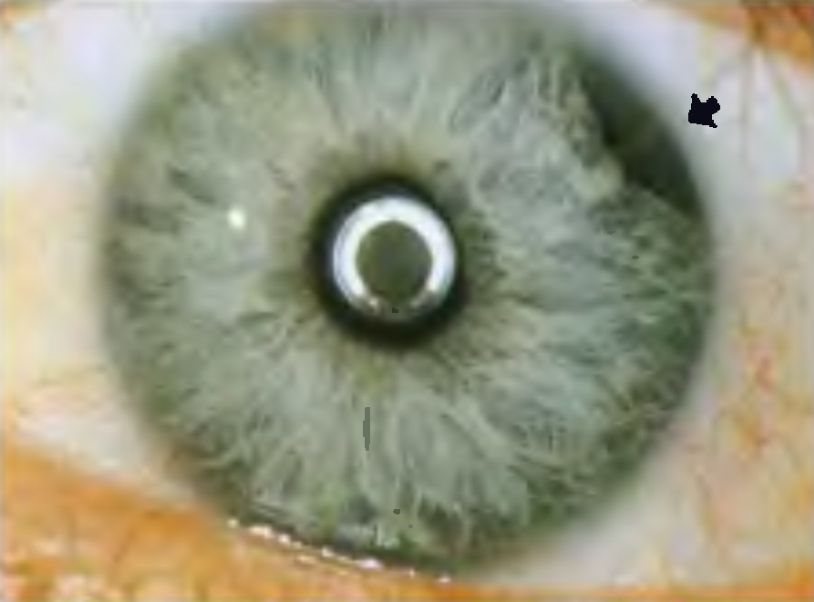
2) What historical features are concerning for intra-ocular foreign body?
Orbital and Intraocular foreign bodies
- Need clinical suspicion
- Hammering, grinding, metalworking, machine operating
- Explosions, firearm use
- Need CT diagnosis
- Treatment:
- Based on optho opinion;
- Inert bodies (plastic, glass, metals) may be left in
- Organic and oxidizing material needs removal
- Need eye shielding
- Need IV ceftazidime
- Need topical erythromycin
- Based on optho opinion;
3) List 4 options for treatment of corneal abrasions
Mechanical Corneal Abrasions
- FB sensation, photophobia, decreased VA
- Pain relief with topical anesthetics diagnose the problem as corneal injury
- Watch for a positive Seidel’s sign – which suggests a corneal perforation
- Treatment
- Full lid eversion and examination!
- Contact lenses shouldn’t be worn until the abrasion is healed (3-5 days)
- Eye patches aren’t needed!
- Cycloplegic prn
- g. Tropicamide
- Topical antibiotics – probably only needed for people who wear contact lenses
- Pseudomonal coverage if contact lens wearer (tobramycin 0.5% 1-2 drops q 4hrs)
- Topical analgesics:
- Ketorolac 0.5% QID
- Diclofenac 0.1% QID
- Tetanus immunization only needed for any “tetanus-prone” injury with dirt and organic matter
- NO cases of tetanus have been documented from simple corneal abrasions
- Symptoms should resolve by 24-72 hrs
Corneal Foreign Bodies
- High risk features for perforation
- Grinding, drilling, saws, hammering –> consider CT orbits
- Treatment
- full eye exam
- Topical anesthetic
- Remove:
- Irrigation, moistened cotton tip applicator
- 18 ga BLUNT needle
- Rust ring:
- Needs 24 hrs to prime and move to the surface of the cornea
- Referral
- Deeply embedded, in the visual axis
Conjunctival Foreign Body
- Same approach as corneal but less risk of affecting vision
- Use topical phenylephrine to help reduce the bleeding on removal
Subconjunctival Hemorrhage
- Common occurrence with valsalva or spontaneously
- Should be PAINLESS, not affecting vision, with no photophobia
- Should not tract into the limbus
- If bilateral:
- Think about bleeding diathesis
- Treatment: cold compresses x 24 hrs
- Resolves in 2-4 weeks
4) Describe the management of traumatic hyphema
Traumatic hyphema
- Due to injury to the blood vessels in the iris or ciliary body
- Amount varies from miniscule (all what can be seen by slit lamp — > full “8 ball”)
- Symptoms:
- Pain / photophobia / dec. VA / mildly elevated IOP
- Management:
- Need admission if:
- >50%, decreased VA, increased IOP, sickle cell disease
- g. “really big, really bad, gonna pop, or patient factors”
- Treatment (if no sickle cell disease)
- Topical beta blocker
- Topical alpha-agonist / carbonic anhydrase inhibitor
- Acetazolamide or IV mannitol
- +/- Cycloplegics and steroids
- At risk for:
- Rebleeding in 2-5 days
- Corneal blood staining
- Glaucoma (due to angle recession)
- Synechia formation
- Those with hemoglobinopathies:
- Sickle cell disease / trait ; thalassemia are at increased risk for complications
- AT high risk for INCREASED IOP
- Need coordinated intensive treatment with opthalmology
- Sickle cell disease / trait ; thalassemia are at increased risk for complications
- >50%, decreased VA, increased IOP, sickle cell disease
- Need admission if:
Traumatic iridocyclitis (uveitis)
- Caused by blunt injury to the globe — > ciliary spasm
- Symptoms:
- Photophobia / deep aching pain
- Signs
- Perilimbal conjunctival injection (ciliary flush)
- Cells in the ant. chamber
- Flare (protein content)
- Non-dilating pupil.
- Direct and consensual photophobia
- Treatment:
- Long acting cycloplegic (homatropine)
- Prednisolone
Traumatic mydriasis and miosis
- Need to rule out altered LOC or cranial nerve defect before a pupillary defect is diagnosed
- Results from small tears in the pupillary muscle
5) What causes the finding of a ‘second pupil’ post-trauma?
Iridodialysis
- Tearing of the iris root from the anterior ciliary body –> leads to second pupil
- Usually occurring after blunt trauma
- Watch for hyphema
- Symptoms: monocular diplopia
- Needs immediate optho. consult;
- Bed rest
- Keep intraocular pressure low
- Eye shielding
6) Describe the physical findings of globe rupture and describe management
Scleral globe rupture
- Occurs in setting of blunt or penetrating trauma
- May be obvious (contents oozing) or subtle
- Symptoms:
- VA / pain
- Signs:
- Bloody chemosis / severe subconjunctival hemorrhage /
- Tear drop pupil
- RAPD / poor VA / no red light reflex
- Do NOT do tonometry
- CT:
- Only 75% sens.
- Treatment:
- Eye shield
- Head of bed > 45 degrees
- NPO
- Antiemetics
- Analgesics
- Antitussives
- Broad spectrum abx:
- Ceftriaxone & gentamicin & vancomycin
7) List 5 indications for ophtho consultation for eyelid lacerations
Laceration of the eyelids
- Need to search for a penetrating injury and foreign body
- Simple superficial lacerations not involving the eyelid margin can be treated in emerg.
- Simple 6-0 / 7-0 interrupted sutures removed in 3-5 days
- Complex lacerations needing referral:
- Lac of the lid margin
- Of the canalicular system (medial eyelid)
- Involving the levator or canthal tendons
- Through orbital septum
- Presence of orbital fat*** = no subcutaneous fat in the eyelids so the fat is likely from a globe injury
- With tissue loss
Conjunctival / corneal / scleral lacerations
- Small superficial conjunctival lacerations = no suturing, heal well,
- Topical antibiotics
- Larger (> 1cm)
- Need repair
- Corneal/scleral lacerations
- Full thickness if:
- Loss of anterior chamber depth, teardrop-shaped pupil, blood in anterior chamber, seidels signs
- Treated:
- As globe rupture with optho. consult
- Full thickness if:
8) Describe diagnosis and treatment for orbital floor fractures
Orbital wall fractures
- The orbital floor is the weakest point = it’s the emergency pressure release to prevent globe injury
- Fracture can lead to entrapment of inferior rectus/oblique muscle; orbital fat or connective tissue
- Signs:
- Enophthalmos, ptosis, diplopia, anesthesia of cheek and upper lip, limitation of upward gaze
- Diagnosis: CT orbits is the preferred test
- Treatment:
- If fracture into an infected sinus:
- Decongestants +/- steroids
- Clavulin
- Ice packs
- Medial orbital wall (enter the ethmoid sinus)
- Signs
- Orbital emphysema and epistaxis
- Diplopia
- Key instructions:
- Don’t blow your nose or sneeze
- Watch for signs of infection
- Watch for double vision or visual loss
- Can be discharged home if:
- No globe rupture
- No visual impairment
- Signs
- If fracture into an infected sinus:
a) List 2 findings on X-ray of orbital floor fracture
- Plain x-ray films have limited utility:
- On x-ray film, the teardrop sign, a bulge extending from the orbit into the maxillary sinus,
- An air-fluid level in the maxillary sinus are indirect signs of orbital floor injury
b) List indications for surgical repair of orbital floor fracture
- Surgery for:
- Persistent diplopia +/- loss of visual acuity
- Cosmetic concerns that persist after 7-10 days when swelling has subsided
- Don’t need “in ER” consultation, can be seen in f/u in 1-2 weeks
- Consider admission and quicker consultation if the fracture extends through an infected sinus
9) Describe the clinical findings of retrobulbar hemorrhage and the steps in performing lateral canthotomy
Retrobulbar hemorrhage
- Causes acute rise in IOP which can compress the optic nerve
- Compression of the Central retinal artery and optic nerve
- Signs
- Proptosis
- Limited EOM
- Visual loss
- Increased IOP
- ***Don’t wait for a CT scan if you are suspicious*****
- Treatment:
- Carbonic anhydrase inhibitor
- Topical beta blockers
- Mannitol 1-2 g/kg
- LATERAL CANTHOTOMY

The procedure:
- Ensure the patient has one of the absolute / relative indications for this procedure
- DIP A CONE
- Informed consent
- Don PPE
- Wash the area with saline
- 1-3 ml 1% lidocaine with epi. Into the lateral canthus (consider light procedural sedation)
- Devascularize with hemostat
- Incise the lateral canthus
- Pull lower lid down and localize the inferior canthal tendon – then cut it with iris scissors
- Reassess, and repeat for the superior canthal tendon if needed

Table copied from: https://www.ncbi.nlm.nih.gov/pubmed/17637149
See:
https://first10em.com/2015/04/01/procedure-lateral-canthotomy/
And
http://webeye.ophth.uiowa.edu/eyeforum/tutorials/lateral-canthotomy-cantholysis.htm
For videos explaining it!
10) List 3 complications of ocular trauma
Post-traumatic corneal ulcers:
- Can develop post-trauma due to bacterial or fungal infection
- Signs: white/gray cornea
- Hypopyon
- Treatment:
- Ophtho referral
- Cycloplegic
- Topical antibiotics
Endophthalmitis
- Infection of the DEEP structures of the eye
- Anterior, posterior, vitreous chambers of the eye
- Symptoms:
- PAIN, and vision loss
- Signs:
- Decreased VA, chemosis, hyperemia, hazy chambers
- Risk factors:
- Blunt globe rupture, penetrating eye injury, foreign bodies, ocular surgery
- Treatment:
- IV ceftazidime, IV vancomycin
- intraocular gentamicin + clindamycin
Sympathetic ophthalmia
- Famous disease: thought to have affected Louis Braille who was blind by age 5!
- http://eyewiki.aao.org/Sympathetic_Ophthalmia
- Inflammation that occurs in the UNINJURED EYE weeks to months after opposite eye injury
- An autoimmune response to the normal uveal tissues
- Symptoms:
- Pain, photophobia, dec. VA
- Treatment:
- Steroids, immunosuppressive agents
This post was uploaded and copyedited by Colin Sedgwick (@colin_sedgwick)

Adam Thomas
Latest posts by Adam Thomas (see all)
- CRACKCast E191 – Weapons of Mass Destruction - July 2, 2018
- CRACKCast E189 – Air Medical Transport - June 25, 2018
- CRACKCast E188 – Emergency Medical Service: Overview and Ground Transport - June 21, 2018
Latest posts by Chris Lipp (see all)
- CRACKCast E191 – Weapons of Mass Destruction - July 2, 2018
- CRACKCast E189 – Air Medical Transport - June 25, 2018
- CRACKCast E188 – Emergency Medical Service: Overview and Ground Transport - June 21, 2018